
Total Knee Replacement
Worn-out knee joints from arthritis, and the knee after a total knee replacement.
Pending Medical Review
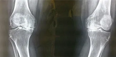
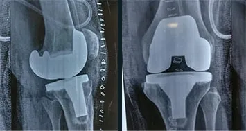
Families come to Dr. Gadkari with painful joints, broken bones and worn-out knees. Here is what some of them wrote afterwards, in their own words, and what their X-rays looked like before and after surgery.
“Your skill, dedication, and compassionate approach have made a remarkable difference in my recovery and overall well-being.”
“His diagnosis is always spot on, he explains things clearly and patiently, and thanks to him we’ve all recovered really well.”
“We found him extremely approachable responding to our queries 24x7, providing practical solutions and not prescribing excessive medication.”
“We consulted Dr Kuldeep, he did not immediately recommend a surgery! Prescribed medication & managed conservatively till the pain subsided after which he decided to get her operated.”
“He is patient, articulate, gives feedback at various stages, takes ownership of the clinical journey and is updated with the latest technology in orthopedics.”
“He had done my surgery very nicely and after two days of surgery only I am walking on my both the legs freely and easily.”
“He explained in detail the diagnosis and recommended treatment and all through with his calm demeanor and pleasing attitude helped keep my mother in law relaxed providing confidence of speedy and complete recovery.”
“He is a serious professional and he made sure he explained me the procedure in detail before my surgery. It made me feel really relaxed, comfortable and at ease.”
“My left wrist mobility is now very good and my wrist feels normal, thanks to the expertise of Dr. Kuldeep.”
“Dr. Kuldeep gave invaluable advice pre and post my mother's total knee replacement surgery. It was very helpful to know each and every step to follow before the surgery, and the recovery milestones to expect.”
“Dr. Kuldeep examined me & assured me that pain can be relieved through proper exercises & medication.”
“I had a metatarsal fracture and he explained me the line of treatment and made it a very comfortable and smooth process.”
“The surgery went smooth, so did the complete recovery process. Full stars to the doctor for his prompt assistance always.”
“In my experience Dr Kuldeep Gadkari is an extraordinary intelligent Doctor with human values.”
“We were extremely satisfied with his consultation n treatment, which we felt as perfect. We rate him as an outstanding orthopedic surgeon in Thane.”
“Always thinks from patient's perspective and suggest what is best for the patient. Listens calmly and explains in detail, allowing patients to make an informed decision.”
“Unlike other doctors, he carefully explains his diagnosis. My interactions with him were reassuring, and like many others, I am glad that I approached him for a treatment.”
“As a person, I found him both calm and amiable. He takes time to explain his diagnosis. I felt he had a sense of empathy for his patients.”
“Always ready with solutions and gives personal attention, which is utmost important in today’s world!!”
“Dr Kulddep carries a lot of expertise and experience with him. Especially good for joint care and replacement.”
“One of the expert,realiable and patient friendly orthopedic surgeon in Thane.”
Short excerpts from Google reviews, quoted word for word. Each links to the original review.
From Dr. Gadkari's own cases. Identifying details have been removed. Every patient's result is different.
Worn-out knee joints from arthritis, and the knee after a total knee replacement.
Pending Medical Review
An earlier hip replacement that needed to be redone, replaced with a longer revision implant.
Pending Medical Review
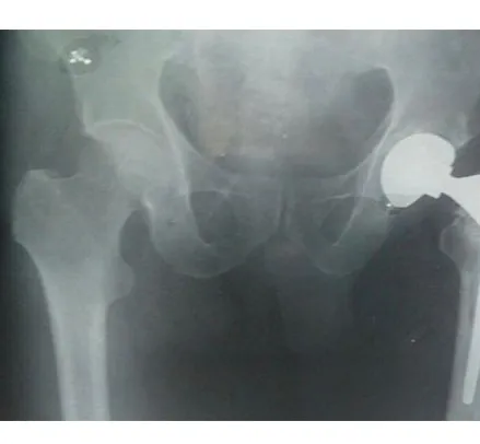
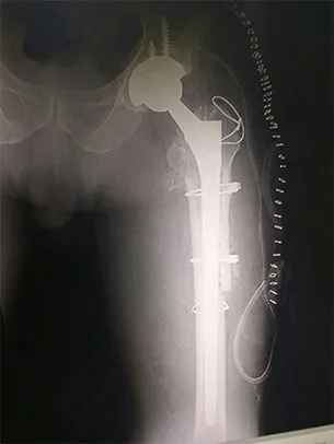
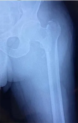
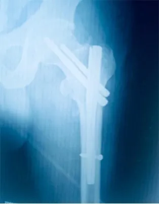
An intertrochanteric (upper thigh bone) hip fracture, fixed with a proximal femoral nail (PFN).
Pending Medical Review
Talk to Dr. Gadkari about your joints. Message the clinic on WhatsApp or call, and they will find a time and place that suits you.